Franklin Diffraction
1 2025-01-22T09:20:29-05:00 George Shaohua Qiao 78e5371050dd5c0e21d36bad469c70d8d4be5464 225 1 "Photo 51" from Rosalind Franklin's diffraction results. plain 2025-01-22T09:20:29-05:00 George Shaohua Qiao 78e5371050dd5c0e21d36bad469c70d8d4be5464This page is referenced by:
-
1
2025-01-22T09:20:26-05:00
Chapter 6: Franklin and Wilkins lay the foundation for solving the structure of DNA
9
plain
2026-07-22T09:57:50-04:00
“The results suggest a helical structure (which must be very closely packed) containing 2, 3 or 4 co-axial nucleic acid chains per helical unit and having the phosphate groups near the outside.”
As we have seen, the story of the discovery that the genetic material is DNA unfolded in Manhattan and Cold Spring Harbor. The next challenge was solving its structure, a sequence of events that took place across the Atlantic in London and Cambridge. Nonetheless, looming over the intense effort to determine the structure of DNA was the towering figure of Linus Pauling at the California Institute of Technology. Pauling was one of the greatest chemists of all time, and his interest in solving the structure helped turn it into a race, as we shall see in the next chapter.
Linus Pauling (1901-1994) won a Nobel Prize in 1954 for his celebrated contributions to chemistry, such as our understanding of electron orbitals, chemical bonding and electronegativity. He also made fundamental contributions to molecular biology in discovering the α-helix and β-sheet structures of proteins. As he explains in the video (click on the icon), Pauling discovered the α-helix simply by model building, which as we will see influenced Watson and Crick in their quest for the structure of DNA. As we will also come to later, he was the first to demonstrate a direct connection between a mutation (sickle cell anemia) and a specific alteration in a protein (hemoglobin). Later in his life, Pauling became a vigorous advocate for a ban on nuclear bomb testing for which he would win a second Nobel Prize, the Peace Prize in 1962. He also advocated for the benefits of mega doses of vitamin C, but this was not, and is not, accepted by the medical community.
Here, we focus on his participation in the story of how the structure of DNA was solved (and return to his impact on the race to solve the structure in the next chapter).
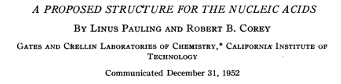
Having only the limited X-ray diffraction data of Astbury and Bell (below) and of CalTech collaborator and crystallographer Robert Corey, Pauling applied his model building approach to predict a structure for DNA. Pauling’s model famously proposed that DNA is triple stranded with the phosphates on the inside and the bases pointing out [L. Pauling and R.B. Corey, “A proposed structure for the nucleic acids” Proc. natn. Acad. Sci. 39:84 (1953)]. Pauling and crystallographer Corey proposed that the peripheral bases could directly interact with and order amino acids:“It is interesting to note that the purine and pyrimidine groups, on the periphery of the molecule, occupy positions such that their hydrogen-bond forming groups are directed radially. This would permit the nucleic acid molecule to interact vigorously with other molecules. …...the 3.4-A x-ray reflection, indicating a similar distance along the axis of the molecule, is approximately the length per residue in a nearly extended polypeptide chain, and accordingly the nucleic acids are, with respect to this dimension, well suited to the ordering of amino-acid residues in a protein.”
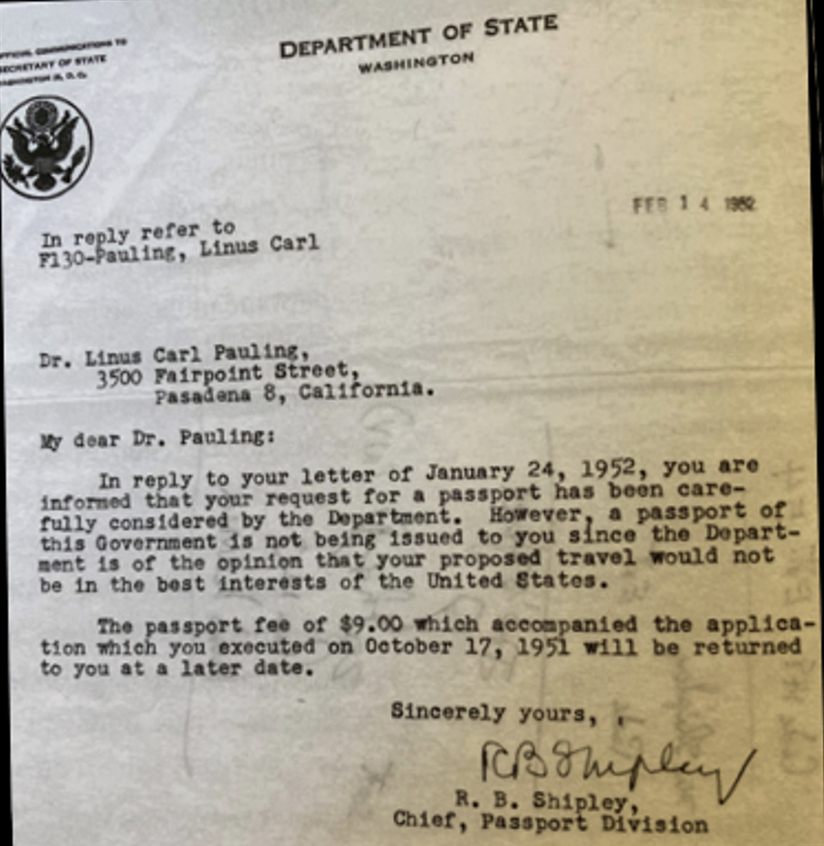
In proposing a triple stranded structure, he failed to consider the contribution of water to the mass of the DNA fibers, and in placing the phosphates on the inside he mistakenly assumed that the closely packed phosphates would be protonated, which would not be the case at physiological pH. He had overlooked that DNA is an acid. Perhaps if he had had access to the higher resolution X-ray diffraction images from the Biophysics Unit of King’s College London, he might have gotten it right. But a planned trip to England in 1952 was blocked by the Department of State because of his political activism (see the letter to the left). Ironically, Pauling had earlier (1948) predicted that the gene would consist of TWO complementary parts:
"If the structure that serves as a template (the gene or virus molecule) consists of, say, two parts, which are themselves complementary in structure, then each of these parts can serve as the mould for the production of a replica of the other part, and the complex of two complementary parts thus can serve as the mould for the production of duplicates of itself."
If he had only remembered his earlier prediction, perhaps he would have realized that DNA had to consist of two complementary strands.
Another clue Pauling overlooked was Chargaff’s Rule that A = T and G = C. According to his son Crelin, Pauling met Chargaff, whom he took a dislike to (Chargaff was famously difficult), on the Queen Mary while sailing across the Atlantic Ocean (video). Years later Pauling pondered whether if he had read his papers instead of being trapped with him on the Queen Mary, he might have taken Chargaff’s publications more seriously.
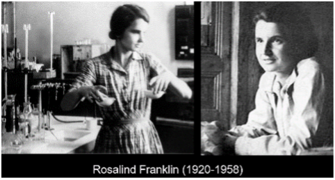
In England, the X-ray structure of DNA was being tackled at the Biophysics Unit of King’s College London, which was under the leadership of Director Sir John Randall. An accomplished X-ray crystallographer, Rosalind Franklin joined the Biophysics Unit in 1951. Maurice Wilkins and his then graduate student Raymond Gosling were already carrying out X-ray diffraction studies of DNA. Nonetheless, Randall wanted Franklin to take over the DNA diffraction work and to supervise Gosling but had not made this clear to Wilkins. This resulted in a tense, combative relationship between Wilkins and Franklin.
Despite the tension, Franklin, now joined by Gosling, made rapid progress. In notes to a lecture she gave at King’s College in 1951, Franklin wrote:“The results suggest a helical structure (which must be very closely packed) containing 2, 3 or 4 co-axial nucleic acid chains per helical unit, and having the phosphate groups near the outside.”
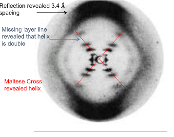
By 1953, she had concluded from her high resolution, X-ray diffraction results, which are famously captured in “Photo 51," which was taken by Gosling (above):- that DNA is a helix (as inferred from the “Maltese Cross” labeled in red)
- that DNA consists of two polynucleotide chains (as inferred from the missing later line)
- that the two chains are not equally spaced.
- that there are 10 residues (nucleotide base pairs) per turn of the double helix with a spacing of 3.4 Å (as inferred from the strong outer reflection)
- that the diameter of the helix is 20 Å
Franklin and Gosling’s contributions were published in 1953 in the journal Nature together with those of Watson and Crick and of Maurice Wilkins and his co-workers. Franklin and Gosling notably wrote:
“Thus, while we do not attempt to offer a complete interpretation of the fibre-diagram of structure B, we may state the following conclusions. The structure is probably helical. The phosphate groups lie on the outside of the structural unit, on a helix of diameter about 20 A. The structural unit probably consists of two co-axial molecules which are not equally spaced along the fibre axis, their mutual displacement being such as to account for the variation of observed intensities of the innermost maxima on the layer lines; if one molecule is displaced from the other by about three-eighths of the fibre-axis period, this would account for the absence of the fourth layer line maxima and the weakness of the sixth. Thus our general ideas are not inconsistent with the model proposed by Watson and Crick in the preceding communication.”
Meanwhile, Wilkins, who had continued his X-ray diffraction studies after Franklin joined the Biophysics Unit, reported in the second of the three back-to-back publications in Nature, that “Such a system of helices (corresponding to a spiral staircase with the core removed) diffracts mainly over a limited angular range, behaving, in fact, like a periodic arrangement of flat plates inclined at a fixed angle to the axis.” We note that Wilkins et al. paid special attention to the broad, biological significance of their findings, investigating the structures from sperm heads, bacteriophage and transforming principle:
Sperm heads. It may be shown that the intensity of the X-ray spectra from crystalline sperm heads is determined by the helical form-function in Fig. 2. Centrifuged trout semen give the same pattern as the dried and rehydrated or washed sperm heads used previously. The sperm head fibre diagram is also given by extracted or synthetic.
Bacteriophage. Centrifuged wet pellets of T2 phage photographed with X-rays while sealed in a cell with mica windows give a diffraction pattern containing the main features of paracrystalline sodium nucleate as distinct from that of crystalline nucleoprotein. This confirms current ideas of phage structure.
Transforming principle (in collaboration with H. Ephrussi-Taylor). Active deoxypentose nucleate allowed to dry at 60 per cent humidity has the same crystalline structure as certain samples of sodium thymonucleate.”Wilkin’s X-ray diffraction image (to the left) was of lower resolution than that of Franklin and Gosling (above) and less revealing of the structure of DNA, though he would share the Nobel Prize with Watson and Crick. Nonetheless, he is to be credited with the insight of focusing on DNA and tackling its structure by crystallography before Franklin arrived at King’s College. However, Wilkins was not the first person to apply X-ray crystallography to study the structure of DNA. The pioneers were William Astbury and Florence Bell (later Sawyer) of the University of Leeds. In 1938 they described nucleotides in DNA as a “pile of pennies,” but they did not interpret their diffraction images as indicating helicity. (As we will see, Crick’s theory of X-ray diffraction pattern for helices had not yet published.)
Returning to Franklin, her fraught relationship with Wilkins and Randall led to her leaving King’s College in 1953 and moving to Birbeck College where she switched her focus to the structure of viruses and was joined by future Nobel Prize winner Aaron Klug. Just three years later Franklin was diagnosed with ovarian cancer, possibly due to excessive exposure to X-rays. She moved in with Crick and his wife and others as she was convalescing following treatments for her cancer. She tragically died at only age 37.